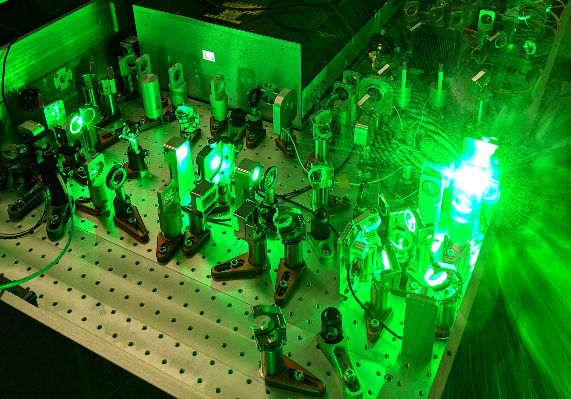
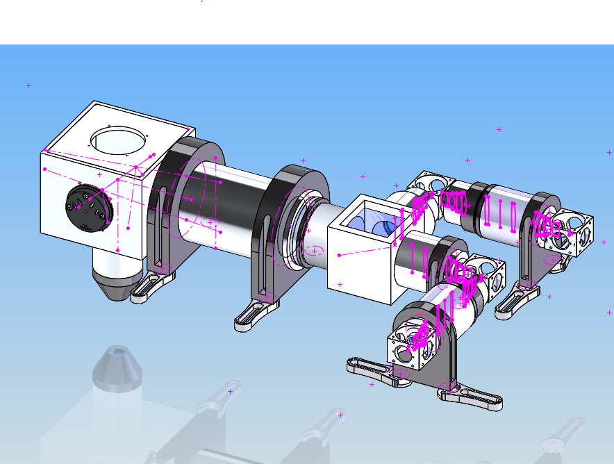
Lab Technology
The usable maximum imaging depth with two-photon microscopy depends on the fluorophore, e.g., for gCaMP6, it is typically about 450–500 μm from the cortical surface. But for far shifted red dyes such as Alexa 680 dextran, an imaging depth of 800 um is routinely achieved. While the mouse visual cortex is only about 750 μm thick, non-rodent mammals have a visual cortex that is 2 mm deep. Thus, much of the circuitry is missed when using two-photon microscopy. For example, layer 4 imaging of the non-rodent neocortex is of paramount importance in understanding cortical computation because it is the layer in which almost all visual cortical receptive field properties emerge de-novo. Just one synapse preceding layer 4 in the primary visual cortex, i.e., in the lateral geniculate nucleus of the thalamus, neurons do not have elongated oriented receptive fields and are not tuned for stereoscopic binocular vision. Therefore, for imaging cortex from 800–2000 μm below the brain surface, our laboratory has setup two microscopes for 3-photon imaging.